Patient Information

Tibialis posterior tendinopathy
Print this page
![]()
Information in this booklet is intended to be used as a guide. It gives you an idea about how tibialis posterior tendinopathy can be managed.
You should remember that every case is different, and symptoms and management can vary from person to person.
Tibialis posterior tendinopathy can be a painful condition affecting the inside of your ankle around your ankle bone. It can lead to a reduction in the arch support around the inside of your foot. This can cause flat feet, as well as swelling and pain in this area.
The condition is often due to an increase in loading, such as more walking or even running. It is not something to be worried about; however, it often does not get better on its own and typically requires some treatment. The first line of treatment includes physiotherapy-led strengthening exercises. Other effective treatments you can complete at home are highlighted in this booklet.
This booklet will provide further information on the condition and then some treatments and exercises to help the process of reducing pain and aiming to return to normal activity.
What is tibialis posterior tendinopathy?
The tibialis posterior is a muscle in your lower leg, around your calf, which helps to point your toes and turn your foot inwards. The muscle also helps to support the natural arch in your foot. The muscle runs down the inside of the lower leg and then travels around the ankle, before attaching to bones underneath the arch of your foot.
A tendon is a tough structure that attaches a muscle to a bone.
Tendinopathy is a term used to describe age-related changes and/or irritation in or around the tendon. It can happen when the tendon struggles to cope with the load or what you are asking it to do.
This can be because the amount it is being used has increased and it has not adjusted, or because it has weakened. This can make activity, especially weight-bearing, difficult and sore.
Due to the tendon and muscle not working very well, it can mean the arch in your foot can drop. This places more stress on other structures involved in maintaining the arch of the foot, such as ligaments, which can also become painful.
Why me?
Tibialis posterior tendinopathy is often due to a recent increase in activity or overuse. With excessive or repetitive loading through activities, such as running and walking, micro-tears may occur within the tendon more quickly than the body can repair them. This can result in tendinopathy and can cause pain, swelling and reduced function.
Sometimes it can be hard to identify why it has happened, and it may have developed more slowly over time.
Anybody can get tibialis posterior tendinopathy, some factors contribute to its development:
Flat feet/shallow arches
age
activity levels
other risk factors include obesity, diabetes and certain inflammatory conditions.
If you already have quite flat arches, then you are more likely to develop symptoms. The condition tends to be more commonly seen in woman over the age of 40, however men can still get symptoms.
Overuse is a very common cause, and people who do a lot of walking or high-impact sports are more likely to experience problems. If the load and impact on the tendon is higher, then you are more likely to develop symptoms.
People who are obese have a higher risk of developing these symptoms.
Symptoms
The symptoms typically come on gradually and can vary in intensity. The symptoms will normally worsen over time and do not normally improve on their own without some form of treatment:
Initially, you may be aware of discomfort and/or swelling behind the ankle and along the instep.
The pain may become worse, particularly with increased weight- bearing.
High-intensity or high-impact activities, such as running, can be very difficult. Some people can have trouble walking or standing for long periods.
You may become increasingly aware of your foot becoming flatter over time.
You may begin to experience pain on the outside of your foot and ankle, as the change in foot position places increased stress between the bones in this part of the foot.
In the later stages, you may find that, on the affected side, you are unable to stand on one leg and raise your heel.
Diagnosis and investigations
Tibialis posterior tendinopathy is diagnosed from the signs and symptoms that you describe. Assessment of the foot, ankle and knee by a healthcare professional may help to inform this diagnosis. If the diagnosis is unclear, an x-ray or ultrasound may be performed to aid the diagnosis.
Blood tests are not used to diagnose tibialis posterior tendinopathy, but they may be used to rule out other contributing factors.
Will it get better?
Your symptoms can often be managed with advice and exercises from your physiotherapist.
Most cases will resolve with conservative treatment within three to 12 months.
Treatment is aimed at:
Reducing pain and inflammation, and promoting healing.
Restoring flexibility and normal movement.
Improving and normalising function.
The tendon and muscle can get stronger, and soft tissues can improve and recover well with the right treatment and advice.
We work with a team of advanced clinical practitioners and orthopaedic consultants who can review your case if symptoms do not improve within an expected period of time. However, steroid injections and surgery are rarely advised for the treatment and management of posterior tibialis tendinopathy.
Management
Tibialis posterior tendinopathy is treated with a variety of different management techniques. Below is a list of ways you can help yourself.
Rest: It is recommended that you avoid excessive standing/walking during recovery. You should stop high-impact activities until your symptoms improve.
Footwear: This can play a major role in recovery. Good quality walking boots that offer support both to the arch of the foot and around the ankle are recommended if walking or on your feet for long periods. Avoid wearing unsupportive footwear such as flip-flops.
Medication: Pain-relieving medication such as paracetamol, or medication to help reduce inflammation, such as ibuprofen, can be helpful. It is best to consult a pharmacist for advice on whether these are suitable for you.
Ice: This will help to reduce the pain and swelling. An ice pack, or ice/small frozen vegetables wrapped in a towel, is ideal (do not apply ice directly to the skin as this may cause an ice burn. Apply for 10 to 20 minutes. This can be repeated several times per day, though leave at least two hours between applications.
Medication for pain control
Controlling your pain allows you to continue to function and carry out your daily activities more comfortably.
Your GP may have already discussed medication to help with your pain and the correct ways to take pain relief. They may recommend that you take it as a short course rather than ‘as and when’ the pain is bad. This often includes paracetamol, co-codamol (Zapain), and non-steroidal anti-inflammatory medication such as ibuprofen. Please always read the instructions before using these products.
Physiotherapy
Specific exercises to rehabilitate and strengthen the tendon can help you to return to your previous levels of activity.
Physiotherapy exercises will be started at a level to suit you and then progressed to ensure the tendon continues to get stronger. Further exercises for the lower limb, including the knee and hip, can also help (see exercises below).
Surgery
Most cases will resolve with conservative (non-surgical) management. However, should the problem persist, surgery may be indicated, but this is rare.
There are several different surgical approaches for repairing a damaged tibialis posterior tendon. There are potential complications from surgery, and recovery takes months. For this reason, surgery is only considered if the symptoms have not improved after six to 12 months of appropriate conservative (non-surgical) treatment.
Orthotics and podiatry
You may be referred by your healthcare professional to see a podiatrist or an orthotist who are specialists in the foot and ankle. These services may offer:
Foot orthoses (insoles): These can help to reduce the stress put on the injured tendon. A podiatrist can provide foot orthoses modified to your specific needs
Ankle brace: This may be required if a level of support is needed that cannot be gained from footwear and foot orthoses alone.
Exercises
The following exercises can be done at home. The number of repetitions is a suggestion. If it is painful, then you can simply reduce how many you do or how often you do them.
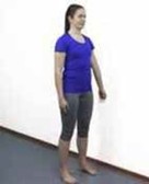
Stand with your feet hip-width apart.
Keeping the toes relaxed and flat on the floor, lift the arches in your feet - you may feel your thighs turning out when doing this.
Hold this position for 10 seconds and then relax.
Reset your feet and repeat the exercise.
Repeat the above 10 times for a total of three sets.
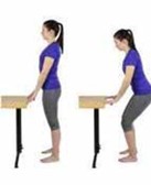
Stand with your feet hip-width apart and your toes slightly pointing outwards (stand in front of a table if necessary).
Keeping your chest up, bend your knees to perform a small squat.
Do not allow the arch in your feet to collapse.
Stop if you feel you are losing your foot position.
Return to the standing position and repeat the exercise.
Repeat the above 10 times for a total of three sets, trying to increase the depth of your squat as you improve.
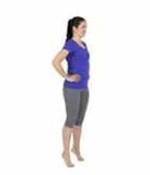
Stand with your feet hip-width apart.
Allowing your knees to bend slightly, lift the heels to stand on your toes while maintaining the position of your feet.
Stop if you feel you are losing your foot position and reset.
You may need to be positioned to hold on to something for balance.
Hold the position for 10 seconds.
Repeat the above 10 times for a total of three sets.
To progress this exercise, put a tennis ball between your inner heels and gently squeeze your heels together to hold the tennis ball as you lift to stand on your toes.
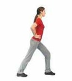
Standing, place the affected foot behind you.
With both feet pointing straight ahead, keep the back heel down and the knee straight.
Lean forward on the good leg to feel a stretch in the calf muscle of the affected leg.
Hold this position for 10 to 15 seconds.
Release and repeat three times.
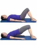
Lie on your back with your knees bent and arms to your side.
Clench your buttock muscles and lift your bottom off the floor or bed.
Keep your back straight, avoiding overarching as you complete the movement.
Hold the position for a few seconds and then lower.
Repeat the above eight to 12 times.
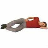
Lie on your side with your bad foot on top of the other.
Bend your knees slightly (as in the picture).
Pull in your lower tummy muscle to activate your core to hold your stomach and back in position so it doesn’t move as you move your leg.
Keeping the feet together, lift the top knee as high as possible (but do not allow yourself to roll back).
Hold the position for a few seconds and then lower slowly.
Repeat the above eight to 12 times.
Your Health Notts
It is always important to consider other factors that can contribute to musculoskeletal problems and may limit your recovery. The most common factors that can affect your health and wellbeing include smoking, alcohol, weight, stress and general fitness.
There is an excellent resource now available to Nottinghamshire County residents that offers information and guidance on:
- Stopping smoking
- Losing weight
- Alcohol reduction
- Increasing physical activity.
Please visit the Your Health Notts website to find out more or self-refer to this service.
Further sources of information
This document is intended for information purposes only and should not replace advice that your relevant health professional would give you.
External websites may be referred to in specific cases. Any external websites are provided for your information and convenience. We cannot accept responsibility for the information found on them.
Patient Experience Team
The Patient Experience Team (PET) is available to help with any of your compliments, concerns or complaints, and will ensure a prompt and efficient service.
Contact Patient Experience.
If you require a full list of references for this leaflet, please email patient.information@sfh-tr.nhs.uk or call 01623 622515, extension 6927.
Approved
October 2025
Review date
October 2027
Document Id
PIL202510-01-TPT
Service / Department
Musculoskeletal (MSK)
Other formats
Patients who would like this information in an alternative format, or need help communicating with us, please contact our patient experience team.